


The Highest Quality, Faster Results & the Lowest Radiation Radiography
Cone Beam Computed Tomography (CBCT), also referred to as CAT scans, is a truly innovative imaging system that provides high-tech information that would otherwise go unseen. The CT-Scan is the most thorough and accurate dental and maxillofacial imaging technology that creates a high-definition and 3D digital model of dental structures, soft tissues, nerve paths, and bony structures in the craniofacial region. The scanner takes a series of still images, then reshuffles them to create 3d images of the bone and soft tissues of your face and mouth.
The Highest Resolution The Precise Imaging Better Diagnosis Predictable outcome
The Modern & Hi-Tech Radiography with Lowest Radiation
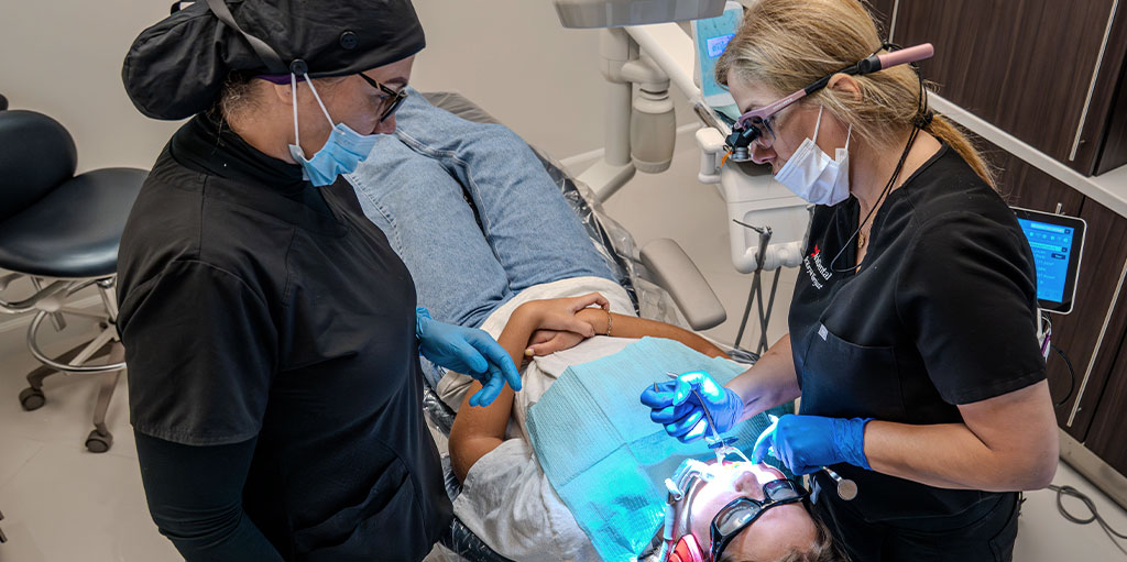
At Aria Dental, Dr. Horiyat has implemented high-fidelity facial 3D Cone Beam CT-Scan to evaluate any ongoing disease, treatment planning, and surgical templating. With immediate access to crystal-clear images, we can diagnose, treat, and educate our patients in a faster and more effective process than ever. 3D dentistry allows us to uncover up to 30 percent more diagnoses which means healthier patients.


How Does It Work?
Low Radiation | Precise | 3D | High Resolution
The 360-degree scan of your head and mouth is fast and painless. Cone beam converts a series of low-dose images into high-resolution views through sophisticated space-age 3D software for designing surgical procedures with greater precision.
The Reasons Your Dentist Orders a Facial CT-Scan?
Dr. Horiyat may recommend high-resolution 3D reconstruction to detect and evaluate certain diseases and pathologies of the jaw, dentition, bony structures of the face, nasal cavity, and sinuses.
Dr. Horiyat regularly utilizes this high-definition technology for many other reasons such as
- To create a surgical template for accurate placement of a dental implant.
- To assess the bone structure and density before implant placement.
- To locate vital structures such as sinus cavities and nerve canals that are critical to identifying for surgical planning.
- To diagnose any pathology in the jaw
She can also use CBCT for treatment planning of orthodontics and dental implants.
Why 3D Cone Beam CT-Scan?
Cone beam CT-Scan can display TMJ disorders, impacted teeth, cracked teeth, hidden infections such as cavitation (jawbone) infection, critical bone and teeth relationships, oral-nasal airway, and mandibular nerve canal. The high-resolution and digitally enhanced pictures provide layers of information that can be separated to view aspects of your mouth, face, and jaws.
3D CT-Scan
CBCT (Cone Beam Computed Tomography) provides a high definition of 3D maxillofacial imaging of dental structures, soft tissues, nerve paths, bony structures, and surrounding vital structures.
Benefits of CBCT
- Precision Placement of Implants in the Bone
- Proper Orientation of the Implants with Its Overlaying Restoration
- Prevention of Injury to Nerves
- Prevent Implant Penetration Into the Sinus
- Selection of the Right Size Implant for Optimal Support
Advantages of 3D Cone Beam CT-Scan
CBCT possessed several advantages over medical CT.
Unlike regular medical CT-Scan, Cone beam CT types create 3D Images of your mouth while cutting the radiation exposure dramatically. The CBCT scan delivers a radiation dose that is 1/10 of a conventional medical CT-Scan.
- Procedure time is as short as 20 seconds
- Patient upright in an open environment … Not being worried about being claustrophobic
- Less effective radiation doses than a traditional cat scan
- Lower cost to the patient, no referral to outside providers
- The major advantage is its ability to image bone and soft tissue simultaneously
Elements of 3D Cone Beam CT-Scan

Impacted Tooth

Implant Placement

Bone Density & Cavitation

Root & Nerve Orientation
Applications of 3D Digital Cone Beam CT-Scan
- Diagnosis of Unhealthy Bone Density, Tooth Root Infection (Cavitation), & Root Canals
- Oral Surgery Analysis
- Surgical Implant Planning for Precise & Safe Placement
- Orthodontic Decisions
- Cephalometric Analysis
- Facial or Dental Trauma
- Diagnosing Temporomandibular Joint Disorder (TMJ/TMD)
- Surgical Planning for Impacted Teeth
- Reconstructive Surgery
- Periodontal Disease
- Detecting, Measuring, & Treating Jaw
- Locating the Origin of Pain or Pathology such as Bone Cancer, Tumors, or Cysts
- Determining Bone Structure & Tooth Orientation
- Fractures
- Airway Studies & Nasal Anatomy: Septum, Turbinate & Sinuses
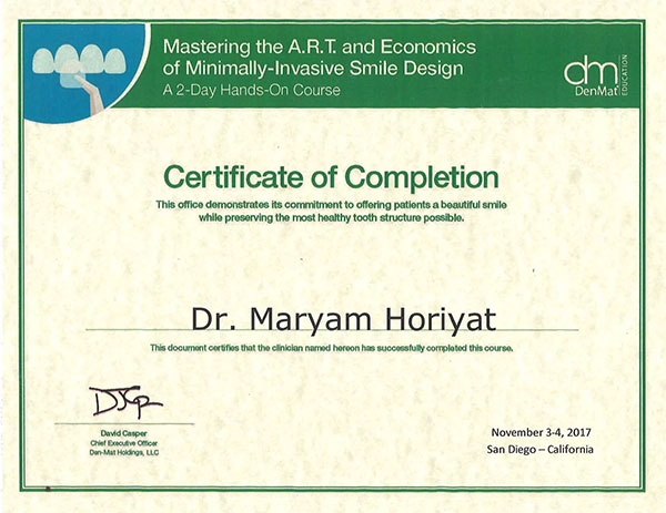
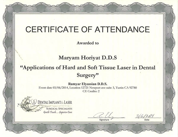
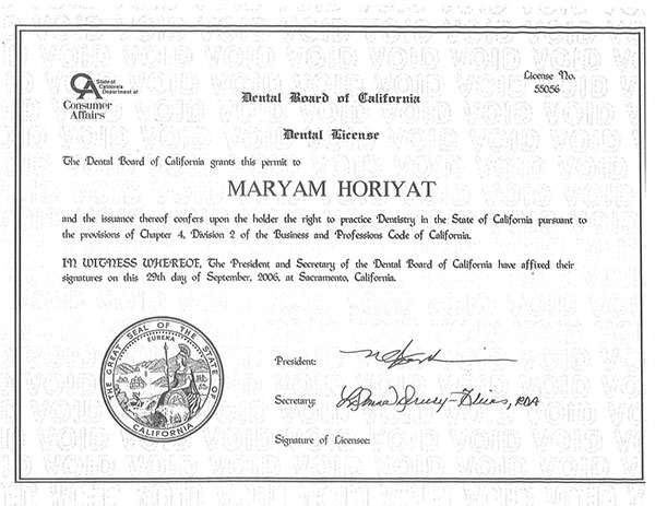
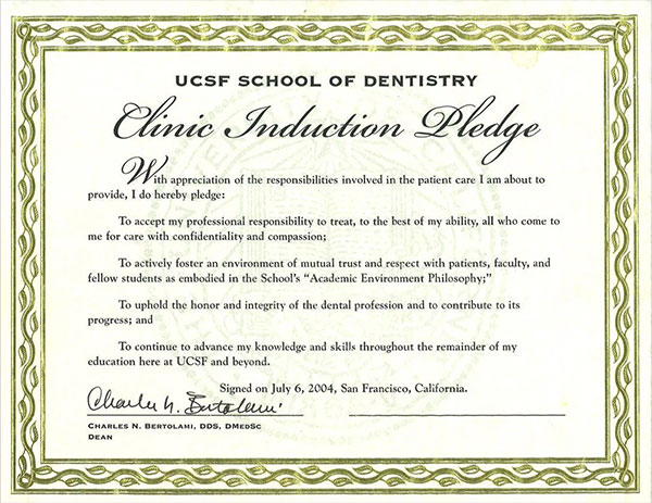
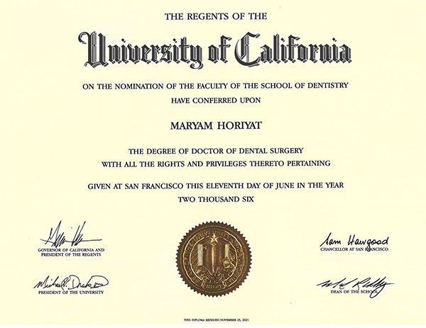
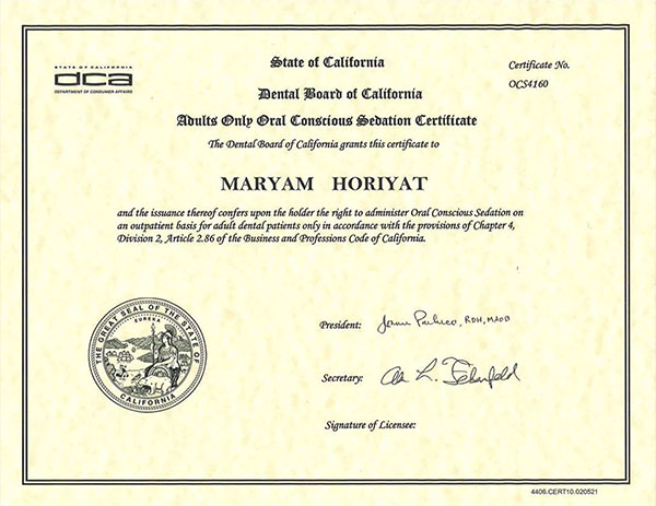
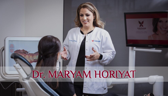
Top 5-Star Advanced Cosmetic & Holistic Dentist
Dr. Maryam Horiyat
in Orange County, CA
OUR Expert Holistic Cosmetic Dentist Experienced Specialists Personal Attention & Gentle Care Holistic & Biomimetic Approaches Outstanding Success Rate Metal-Free Implant Materials Comfortable Sedation Options Modern & Innovative Technologies
OUR Expert Holistic Cosmetic Dentist Experienced Specialists Personal Attention & Gentle Care Holistic, Integrative, & Biomimetic Approaches Outstanding Success Rate & Highest Standards Metal-Free Implant Materials from Highly Reputable Companies Comfortable Sedation Options Modern & Innovative Technologies & Techniques