







A mucocele is a harmless, fluid-filled bump that forms when a tiny salivary gland in the lip, cheek or tongue is injured and saliva leaks into the surrounding tissue. It usually looks like a soft, bluish or clear blister on the inside of the lower lip. Many heal on their own, while persistent ones are removed in a short in-office procedure.
You usually notice it with your tongue first: a smooth, squishy bump inside your lip that was not there last week. It barely hurts, yet it is impossible to ignore. Then it disappears, only to return days later.
If that sounds familiar, you are probably dealing with a mucocele, one of the most common soft-tissue lesions dentists see. This guide explains why a mucocele forms, how to tell it apart from other lip bumps, and what each treatment involves.
Key takeaways
- A mucocele is a benign mucous cyst caused by a damaged or blocked minor salivary gland, most often after lip biting.
- The inner surface of the lower lip is by far the most common location.
- Small, new lesions sometimes heal on their own within days to weeks.
- A mucocele that keeps returning or lasts more than a few weeks is best evaluated by a dentist and usually removed along with the gland that feeds it.
- Popping or cutting a mucocele at home raises the risk of infection, tissue damage and recurrence.
What Is a Mucocele? Understanding Oral Mucous Cysts
Your mouth contains hundreds of minor salivary glands, each far smaller than a grain of rice. They sit beneath the lining of the lips, cheeks, palate and tongue and release saliva through tiny ducts.
A mucocele forms when one of those ducts is damaged or blocked. Instead of flowing into the mouth, mucus-rich saliva pools in the soft tissue and creates a dome-shaped swelling. The National Library of Medicine’s MedlinePlus medical encyclopedia describes this kind of oral mucous cyst as a painless, thin sac filled with clear fluid, and notes that the lesions on the lips, inner cheeks and underside of the tongue are the ones called mucoceles.
You may also hear it called a mucous cyst, a mucous extravasation cyst or a mucous retention cyst. These names describe the same family of salivary gland lesions.
Extravasation vs. Retention Mucoceles: The Two Types
According to the clinical review Mucocele and Ranula in StatPearls, published through the National Center for Biotechnology Information, every mucocele falls into one of two types:
- Extravasation mucocele. This is the common type. A duct is torn, usually by a bite or another injury, and saliva spills into the surrounding connective tissue. Because the pooled fluid has no true lining, it is technically a “pseudocyst.” This type is typical in children, teens and young adults and is strongly linked to the lower lip.
- Retention mucocele. This less common type forms when a duct becomes narrowed or plugged, for example by a tiny salivary stone or scar tissue. Saliva backs up inside a duct that has a true lining. Retention cysts tend to appear in older adults and are more likely to occur in places other than the lower lip.
Mucocele vs. Ranula: Where the Swelling Appears Matters
When a similar mucus-filled swelling forms on the floor of the mouth, beneath the tongue, it is called a ranula. A ranula usually comes from the larger sublingual salivary gland, can grow larger than a lip mucocele, and occasionally extends into the neck. It usually needs an oral surgeon, so not every swelling in the mouth should be treated the same way.
Mucocele Symptoms: How to Recognize the Bump on Your Lip
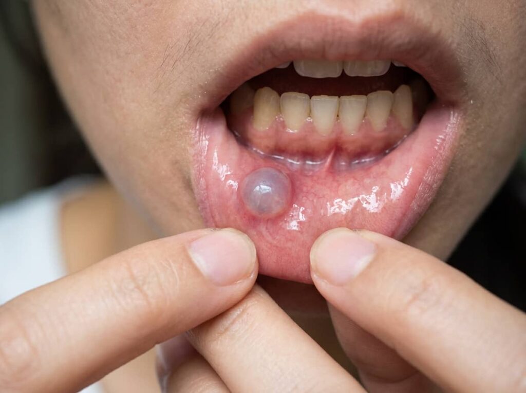
A typical lip mucocele has a recognizable appearance:
- A soft, round, dome-shaped swelling on the inside of the lip
- A translucent, bluish or pinkish color, sometimes resembling a water blister
- A smooth surface that feels fluid-filled when pressed with the tongue
- A size that usually ranges from a few millimeters up to about one centimeter
- Little or no pain, although it can feel awkward or get caught between the teeth
- A tendency to shrink, burst and release a sticky, clear fluid, then fill up again days later
That cycle of swelling, rupture and refilling is one of the most telling signs. If the damaged gland stays active, the fluid collects again.
Superficial vs. Deep Mucoceles
Not every mucocele looks the same. A superficial mucocele sits just beneath the surface as a small, clear blister, often on the soft palate or back of the cheeks, and usually bursts within a day or two. A deep mucocele forms further down, looks pinker, feels firmer and lasts longer. Because it can resemble other lumps, a deep mucocele is more likely to need a professional diagnosis.
What Causes a Mucocele on the Lip?
Most cases trace back to minor trauma that damages a salivary duct. Common triggers include:
- Lip biting or chewing, whether accidental while eating or a stress habit
- Lip sucking, especially pulling the lower lip between the teeth
- A blow to the mouth during sports, a fall or play
- Sharp or chipped teeth, rough fillings or crowns that rub the lip repeatedly
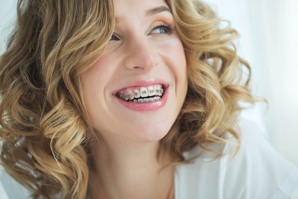
- Orthodontic brackets and wires that press against the inner lip
- Lip and tongue piercings, which irritate the gland tissue around the jewelry
- Teeth grinding or clenching (bruxism), which can go hand in hand with lip and cheek trauma
- Duct blockage from a small salivary stone or scarring, more typical of retention cysts
The lower lip takes the brunt of this trauma because of where it sits relative to the teeth. A 24-year review of 173 cases published in BMC Oral Health found that about 78% occurred on the lower lip, and every one of those lower-lip cases had a history of trauma. The upper lip, by contrast, is a rare location for a mucocele.
Who Is Most Likely to Get a Mucocele?
Anyone can develop a mucocele, but certain groups see them more often:
- Children, teens and young adults, the peak age group in most studies
- People with a habit of biting, sucking or chewing the lip
- Patients wearing braces or other orthodontic appliances
- People with oral piercings
- Anyone with a sharp tooth edge, a broken filling or a bite that traps the lip
A mucocele is not contagious, it is not caused by poor hygiene, and it is not a sign of cancer. It is a mechanical problem with a very small gland.
Mucocele or Something Else? Other Lip Bumps to Rule Out
Several conditions can look similar at first glance:
| Condition | Typical appearance | Key clue |
| Mucocele | Soft, bluish or clear, dome-shaped; may burst and refill | Often follows lip biting; lower lip most common |
| Irritation fibroma | Firm, pink, smooth lump that does not change size | Solid, not fluid-filled; does not drain |
| Hemangioma or vascular lesion | Bluish to purple; may blanch when pressed | Made of blood vessels rather than mucus |
| Canker sore (aphthous ulcer) | Painful, round ulcer with a white or yellow center | An open sore rather than a raised blister |
| Cold sore (herpes labialis) | Cluster of small, painful blisters on the outer lip edge | Tingling beforehand; forms on skin, not inside the lip |
| Salivary gland tumor | Firm, painless, slowly growing lump | More common on the upper lip; does not cycle in size |
| Lipoma | Soft, yellowish, rubbery lump | Fatty tissue; slow, steady growth |
Location is an important clue. A firm, persistent lump on the upper lip deserves a careful evaluation, because a salivary gland tumor is more likely to appear there than a mucocele is.
How Dentists Diagnose a Mucocele
In most cases, a mucocele is diagnosed with a simple clinical exam. Your dentist will:
- Check the size, color, texture and location of the bump
- Feel whether it is fluid-filled or solid
- Ask how long it has been present and whether it has burst
- Ask about lip biting, injuries, piercings, braces and clenching
- Look for sharp tooth edges or restorations that could be the trigger
Imaging is rarely needed, except for large swellings under the tongue or unusual presentations.
When a Biopsy Is Recommended
When a mucocele is removed, the tissue is typically sent to an oral pathology laboratory. This is routine, and it confirms the diagnosis. Laboratory examination is especially important when:
- There is no clear history of trauma
- The lump is firm, growing steadily or larger than expected
- It appears on the upper lip or another unusual location
- It does not follow the classic swell-and-rupture pattern
This step simply ensures that nothing resembling a mucocele is actually something else.

Mucocele Treatment Options: From Watchful Waiting to Removal
The right mucocele treatment depends on size, location, duration, how often it recurs, and the patient’s age and comfort level.
1. Watchful Waiting for Small, New Lesions
MedlinePlus notes that a mucous cyst can often be left alone and may rupture on its own. A small superficial mucocele in particular sometimes heals within days to a few weeks, especially once the habit or irritation that caused it stops. During this period:
- Avoid biting, sucking or poking the area
- Rinse gently with warm salt water after meals
- Skip very spicy, acidic or crunchy foods that irritate the spot
- Keep brushing and cleaning between your teeth as usual
If the bump is still present after several weeks, or keeps returning, waiting is no longer the best strategy.
2. Surgical Excision of the Mucocele and Gland
Conventional surgical removal is the most widely used treatment for a persistent mucocele. After numbing the area with local anesthetic, the dentist or oral surgeon removes the swelling along with the minor salivary gland tissue that feeds it. Removing the feeding gland is the key to preventing recurrence. The site is closed with a few small stitches, and the tissue is sent for examination. For a typical lip lesion, the procedure usually takes well under an hour.
3. Laser Removal
Soft-tissue lasers, such as CO2 and diode lasers, can remove a mucocele while sealing small blood vessels as they work. Potential benefits include minimal bleeding, little or no need for stitches in many cases, and a comfortable recovery. A 2023 case report in the Canadian Medical Association Journal noted that CO2 laser treatment and cryotherapy can be effective with minimal scarring. Laser treatment is often well suited to small and medium lesions and to patients, including children, who may find a scalpel procedure intimidating.
4. Micro-Marsupialization
This conservative technique, used frequently in pediatric dentistry, involves passing a suture through the dome of the mucocele and leaving it in place for about a week. The suture creates new drainage channels, so fluid can escape while the area heals. It avoids cutting tissue, but recurrence is more likely than with full excision, so it tends to be reserved for selected cases.
5. Cryotherapy and Corticosteroid Injections
Cryotherapy freezes the lesion so that it sloughs away, and MedlinePlus lists it alongside laser treatment as a removal option. Intralesional corticosteroid injections are another nonsurgical choice some clinicians use, particularly for a recurrent mucocele or multiple lesions. Both have more limited supporting research than excision, and the choice depends on the individual case.
| Treatment | Best suited for | Main advantage | Main limitation |
| Watchful waiting | Small, new, superficial lesions | No procedure needed | Many lesions recur if the gland stays damaged |
| Surgical excision | Persistent or recurrent lesions | Removes the feeding gland; tissue can be examined | Stitches and a short healing period |
| Laser removal | Small to medium lesions; anxious patients | Minimal bleeding; often no stitches | Equipment and training vary by office |
| Micro-marsupialization | Children; selected small lesions | No cutting of tissue | Higher chance of recurrence |
| Cryotherapy or steroid injection | Selected or multiple lesions | Nonsurgical | Less research; may need repeat sessions |
Why You Should Never Pop a Mucocele at Home
It is tempting to drain the bump with a needle. Resist that urge. MedlinePlus specifically advises against opening the sac at home, because doing so can introduce infection and damage nearby tissue. There is also a practical problem: draining the fluid does nothing to fix the injured gland underneath. In most cases the mucocele simply refills, and repeated self-treatment can create scar tissue that makes a later removal more complicated.
If a mucocele bursts on its own, keep the area clean with saltwater rinses. If it comes back, book a professional evaluation.
Mucocele Removal Recovery: What to Expect After Treatment
Recovery after mucocele removal is usually straightforward. General expectations include:
- First 24 to 48 hours: mild tenderness and swelling, managed with a cold compress on the outside of the lip and an over-the-counter pain reliever taken as directed on the label
- First week: soft foods; avoid spicy, salty and acidic foods; no biting the lip or pulling it down to look at the site
- Stitches: dissolvable sutures typically fall out on their own, while non-dissolving stitches are usually removed about a week after the procedure
- Hygiene: continue brushing carefully, and use warm saltwater rinses as recommended
- Full healing: the lip lining generally heals within one to two weeks, although minor firmness at the site can take longer to settle
Some patients notice temporary numbness in a small area of the lip, because tiny nerve branches run close to the surface. Contact the office promptly if you notice increasing swelling, pus, a fever, or a new bump near the site.
Can a Mucocele Come Back? Preventing Recurrence
A mucocele can return, especially when the gland that produced it is not completely removed, when nearby glands are injured during treatment, or when the habit that caused the first one continues. Recurrence is uncommon after complete excision, although published reports suggest it may be somewhat more frequent in younger patients.
To lower the chance of a repeat mucocele:
- Break the habit. Notice when you bite or suck your lip, especially during stress or concentration.
- Fix sharp edges. A chipped tooth, rough filling or worn crown that rubs the lip should be smoothed or repaired.
- Protect against grinding. If clenching or grinding is involved, a custom night guard can reduce soft-tissue trauma.
- Use orthodontic wax. Patients with braces can place wax over brackets that press against the lip.
- Reconsider piercings. Oral jewelry that repeatedly irritates the same area keeps the cycle going.
- Wear a mouthguard for sports. A properly fitted guard protects lips and teeth from impact.

Mucoceles in Children: A Guide for Parents
Because children often bite their lips, fall and play contact sports, a mucocele is one of the more common oral lumps in young patients. It is very unlikely to be serious.
Helpful steps for parents:
- Take a photo of the bump so you can track changes in size and color
- Discourage poking, biting or squeezing the area
- Schedule a dental visit if the bump lasts more than two to three weeks, keeps returning, or interferes with eating or speaking
- Ask about gentle treatment options such as laser removal or micro-marsupialization, which tend to be well tolerated by younger patients
How Aria Dental Care Approaches Mucocele Evaluation in Mission Viejo
At Aria Dental Care, a mucocele is treated as more than a bump to remove. The goal is to confirm the diagnosis, treat the lesion in the least invasive way that is still effective, and find out why it formed so it does not return.
Dr. Maryam Horiyat, DDS, earned her dental degree at UCSF and is accredited by the International Academy of Oral Medicine and Toxicology (AIAOMT) and certified by the International Academy of Biological Dentistry and Medicine (CIABDM). Her biological dentistry philosophy emphasizes conservative treatment, biocompatible materials and attention to how the mouth connects with overall health.
A mucocele evaluation at Aria Dental Care typically includes:
- A thorough oral exam of the lesion, surrounding tissue, teeth and bite
- A review of habits and triggers, including lip biting, clenching, braces and piercings
- A clear discussion of options, from monitoring to removal, with honest guidance on recurrence risk
- Minimally invasive removal when appropriate, which may include laser dentistry for suitable lesions, or referral to an oral surgeon for larger or deeper cases such as a ranula
- Laboratory confirmation of removed tissue to rule out other conditions
- Comfort options such as sedation dentistry for anxious patients
- Prevention planning, such as smoothing a rough tooth edge, a night guard for grinding, or coordinating with orthodontic care when brackets are part of the problem
Patients visit from Mission Viejo, Laguna Niguel, Rancho Santa Margarita, Irvine and across Orange County. Learn more on the holistic dentist in Mission Viejo page.
Have a Bump on Your Lip? Get Clear Answers
A mucocele can be annoying and a little worrying, but it is almost always a simple, treatable problem. Give a small new bump a couple of weeks. If it lingers, returns or looks unusual, a quick exam brings peace of mind.
Aria Dental Care is located at 27652 Crown Valley Pkwy, Mission Viejo, CA 92691. Call (949) 364-9600 or request an appointment online to have your lip bump evaluated by Dr. Maryam Horiyat.