




“Your gums are receding, and you may need a graft.” For many patients, that sentence lands with a jolt. The words sound surgical, technical, and a little scary. In reality, gingival recession surgery is one of the most common and well-studied procedures in periodontal care, and once it is explained in everyday language, most people find it far less intimidating than they expected.
This guide walks through what gingival recession surgery is, why gums recede in the first place, how the main techniques compare, what recovery is really like, and how the biological, whole-body philosophy at Aria Dental Care shapes every treatment plan.
Quick answer: Gingival recession surgery is a family of periodontal plastic surgery procedures that rebuild gum tissue that has pulled away from the teeth. Using either nearby gum tissue or graft tissue, the surgeon covers exposed tooth roots, thickens thin gums, reduces sensitivity, and helps protect the tooth from further recession, root decay, and bone loss.
What Is Gingival Recession Surgery, in Simple Terms?
Think of your gums as a protective collar that hugs each tooth. When part of that collar wears away, the tooth root underneath is left uncovered. Gingival recession surgery restores the collar. The surgeon either gently moves healthy gum tissue into place or adds a thin piece of graft tissue, then secures it so it heals onto the root surface.
You may hear the procedure called by several names:
- Gum graft surgery or gum grafting
- Soft tissue grafting
- Root coverage surgery
- Periodontal plastic surgery
- Gingival grafting
All of these describe the same goal: putting healthy, protective gum tissue back where it belongs.
Understanding Gum Recession Before Surgery
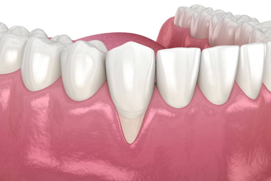
What Happens When Gums Recede
In a healthy mouth, the gum line sits just above the point where the enamel-covered crown meets the root. Gingival recession occurs when the gum margin shifts toward the root and exposes it. The StatPearls clinical reference published by the National Library of Medicine describes recession as a condition frequently seen in general dental practice, particularly in patients with gum disease, shaped by both changeable and unchangeable risk factors.
Why does that exposure matter? The root has no enamel. It is covered by cementum, a softer layer, with dentin beneath that contains tiny channels leading toward the nerve. As a result, exposed roots often react to cold drinks, sweets, and even brushing. They also decay and wear down more easily than enamel. Because lost gum tissue does not grow back by itself, recession usually stays the same or slowly worsens unless the cause is addressed.
How Common Receding Gums Are
Gum recession is remarkably common. A systematic review and meta-analysis indexed on PubMed, which combined 15 population studies and more than 37,000 participants, found that over two-thirds of people worldwide have some degree of recession. Most cases are mild and never need surgery, which is why a careful evaluation matters so much.
Common Causes of Receding Gums
Recession is usually the result of several factors working together:
- Gum disease: Periodontal infection breaks down the tissue and bone that hold the gum in place. Our article on whether gum disease can be dangerous to your overall health explains why this cause is always checked first.
- Brushing too hard: Firm bristles and a back-and-forth scrubbing motion slowly wear gum tissue away.
- Thin gum tissue: Some people are born with delicate gums, known as a thin phenotype, that recede more easily.
- Tooth position: Teeth that sit outside the natural bone envelope, including some teeth moved during orthodontic treatment, may have very little bone and gum covering the root.
- Clenching, grinding, and bite imbalance: Heavy forces are considered a possible contributor, although research on this link is mixed. A custom nightguard for teeth grinding or occlusal bite correction may be part of a complete plan.
- Tobacco and vaping: Both reduce blood flow and slow healing in gum tissue.
- Piercings and a tight frenum: Jewelry or a lip attachment that pulls on the gum line can cause recession in one spot.
- Dental work at the gum line: Poorly fitting crowns, fillings, or partial dentures can irritate tissue over time.
From a holistic standpoint, chronic inflammation, nutrition, stress, and the balance of the oral microbiome also deserve attention, because each influences how well gums hold up and heal.
When Is Gingival Recession Surgery Recommended?
Not every receding gum needs surgery. According to the StatPearls reference above, surgical correction is worth considering when a patient is concerned about appearance or sensitivity, when recession is actively getting worse, or when a tooth with thin or insufficient gum tissue is about to be moved orthodontically. The same source groups surgical options into two purposes: covering exposed roots and adding or thickening firm, keratinized gum tissue.
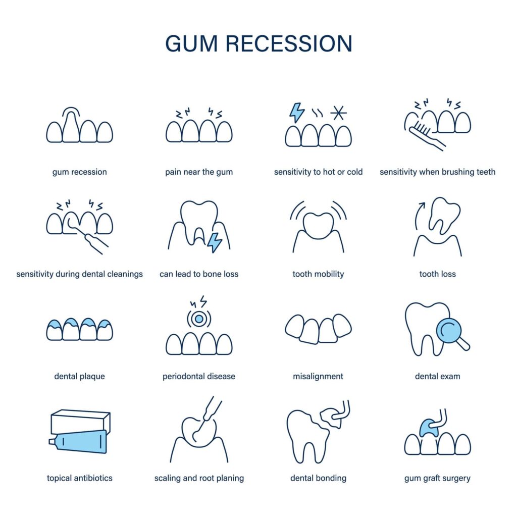
Gingival recession surgery may be worth discussing with your dentist if you notice:
- Teeth that look longer than they once did
- Sensitivity near the gum line that desensitizing toothpaste no longer controls
- A notch, groove, or color change where the gum meets the tooth
- Recession that measures deeper at each checkup
- Gums that look thin or almost see-through over the root
- Planned crowns, veneers, implants, or orthodontics near a thin gum area
How Your Dentist Evaluates Gum Recession
Measuring the Recession
A good plan starts with good measurements. Your dentist records how deep and wide each area of recession is, checks pocket depths around the teeth, evaluates how thick and wide the firm gum tissue is, and reviews X-rays to see bone levels between the teeth. When more detail is needed, a 3D cone beam CT scan and other modern diagnostic technology can reveal bone thickness that standard X-rays miss.
The RT1, RT2, and RT3 Classification
Many clinicians use a system introduced by Cairo and colleagues in 2011 and adopted in the 2017 World Workshop classification of periodontal conditions. It looks at whether attachment between the teeth is still intact, because that largely predicts how much root can be covered:
- RT1: No attachment loss between the teeth. These cases have the best chance of complete root coverage.
- RT2: Some attachment loss between the teeth, but no more than on the front of the root. Partial or full coverage is often possible.
- RT3: Attachment loss between the teeth is greater than on the front of the root. Full coverage is usually not realistic, so the goal becomes stabilizing and protecting the tooth.
The older four-class Miller system follows similar logic and is still mentioned in many offices. Gum thickness matters too: thick tissue heals predictably and resists future recession, while thin tissue is fragile, so some procedures focus mainly on thickening the gum.
The Main Types of Gingival Recession Surgery
The best technique depends on your recession type, the number of teeth involved, the thickness of nearby tissue, how visible the area is when you smile, and your comfort with different graft materials.
Connective Tissue Graft
The connective tissue graft is the most widely used procedure for root coverage and is often described in the research literature as the benchmark technique. The surgeon opens a small flap on the roof of the mouth, removes a thin layer of connective tissue from beneath the surface, and closes the area. That tissue is then placed under the gum at the recession site and held with fine sutures.
As the American Academy of Periodontology explains, gum graft surgery uses tissue from the palate or another donor source to cover the exposed root, helping prevent additional recession and bone loss, and it can treat one tooth or several. Surgeons often pair connective tissue grafts with a coronally advanced flap or a tunnel technique for a natural blend.
Free Gingival Graft
A free gingival graft is also taken from the palate, but it includes the outer surface layer of tissue. It is excellent for building a thick, durable band of firm gum, which makes it popular for lower front teeth with very thin tissue. Because it can heal slightly lighter in color, it is used less often in the most visible smile areas.
Pedicle Graft and Coronally Advanced Flap
If there is enough healthy gum next to the recession, the surgeon can use it directly. In a lateral pedicle graft, neighboring tissue stays attached on one side and is slid over the root. In a coronally advanced flap, the existing gum is released and moved toward the crown. Because the tissue keeps its own blood supply, healing tends to be reliable.
Donor and Collagen Graft Materials
For patients who would rather avoid a wound on the palate, alternatives include processed donor tissue from accredited human tissue banks (acellular dermal matrix), collagen matrices of animal origin, and enamel matrix proteins applied as a gel. These can reduce discomfort and are helpful when many teeth need treatment. Depending on the case, however, the patient’s own tissue may still provide the most predictable thickness. A biological dentist will explain the origin of each material so you can choose with confidence.
Pinhole Surgical Technique
The Pinhole Surgical Technique, developed by Dr. John Chao, is a minimally invasive option. Through a tiny opening, the clinician loosens the gum with specialized instruments, slides it over the exposed roots, and places small collagen strips to help hold it in position. There is no palate wound, and several teeth can be treated in one visit. Its long-term research base is smaller than that of connective tissue grafting, and thin tissue may still need thickening, so careful case selection matters. Our gum recession treatment page covers the options in more detail.
L-PRF and Laser-Assisted Care
Leukocyte- and platelet-rich fibrin (L-PRF) is created by spinning a small sample of the patient’s own blood into a soft, natural membrane. Because it comes from your own body, it fits a biocompatible philosophy, and Aria Dental Care incorporates L-PRF into its surgical protocols where appropriate. Research on L-PRF for root coverage is still evolving, and it is generally used as a helpful adjunct rather than a substitute for grafting in every case.
Laser dentistry plays a supporting role. LANAP laser gum therapy is designed to treat active periodontal disease, one of the main drivers of recession. It does not cover exposed roots on its own, but controlling infection is often a necessary first step before a graft can succeed.
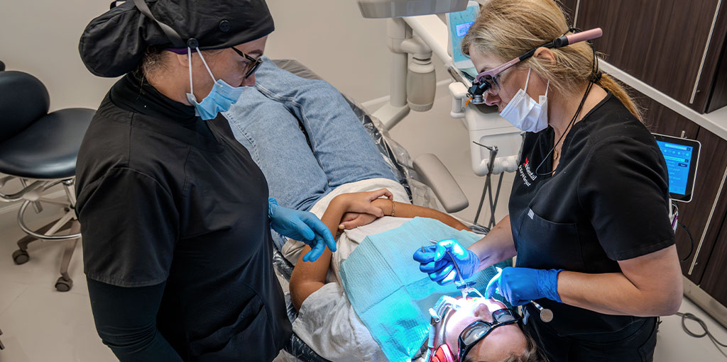
What to Expect on Surgery Day
- Consultation and planning: Your dentist reviews measurements, photos, X-rays, medical history, and medications, then discusses technique and material choices with you.
- A healthy starting point: Inflammation is calmed beforehand, often through a holistic dental cleaning or periodontal gum treatment.
- Comfort first: The area is completely numbed. If you feel anxious, ask about sedation dentistry.
- Preparing the root: The exposed root surface is gently cleaned and smoothed so new tissue can attach.
- Placing the tissue: The graft or repositioned gum is positioned and secured with very fine sutures.
- Protecting the donor area: If palate tissue was used, a dressing or thin clear stent may cover it.
- Going home: Most patients leave the same day with written aftercare instructions.
Recovery After Gum Graft Surgery
The First Few Days
Mild to moderate soreness is normal once the numbness wears off, and swelling usually peaks around day two or three. Cold compresses and rest help. Avoid spitting forcefully, drinking through a straw, or stretching your lip to check the site.
The First Two Weeks
Stick with soft, cool, or lukewarm foods such as yogurt, eggs, mild soups, and tender cooked vegetables. Skip crunchy, spicy, or very hot foods. You will usually avoid brushing the treated area until your dentist clears it, using a gentle rinse instead. Sutures typically dissolve or are removed within one to two weeks, and many patients find the palate more tender than the graft site.
The Following Months
Gentle brushing returns with an extra-soft brush. The new tissue gradually matches the color and texture of nearby gums, and the final gum line generally settles within a few months. Follow-up visits confirm that everything has attached well.
Benefits and Risks of Gingival Recession Surgery
Benefits
- Protects exposed roots from decay and wear
- Reduces or eliminates sensitivity for many patients
- Thickens thin gums, lowering the chance of future recession
- Creates a more even, natural-looking smile
- Builds a stronger foundation before crowns, veneers, implants, or orthodontics
Risks
- Temporary swelling, bruising, or minor bleeding
- Soreness at the palate donor site
- Infection, which is uncommon with proper aftercare
- Partial graft loss or incomplete root coverage
- Small differences in color or texture
- Recurrence if the original cause is not corrected
Careful planning, precise technique, and regular follow-up keep these risks low.
Are You a Candidate for Gum Grafting?
Strong candidates usually have gum disease under control, gentle and consistent home care, stable general health, a willingness to avoid tobacco while healing, and expectations that match their recession type.
Treatment may be delayed if periodontitis is still active, if a condition such as diabetes is poorly controlled, or if medications like blood thinners need coordination with a physician. Mild, stable recession without symptoms may simply be monitored while brushing habits and bite forces are corrected.
Gingival Recession Surgery Cost and Insurance
The cost of gingival recession surgery depends on the number of teeth treated, the technique, whether donor or collagen materials are used, adjuncts such as L-PRF, sedation, and any gum treatment needed first. Many dental plans cover part of gum grafting when it is medically necessary rather than purely cosmetic, though coverage varies.
At Aria Dental Care, you receive a clear written estimate before treatment begins. Our team can help with insurance verification and explain available payment and financing options.
The Aria Dental Care Approach: Biological, Conservative, Whole-Body
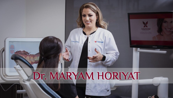
At Aria Dental Care, receding gums are treated as a signal worth understanding, not simply a gap to fill. Dr. Maryam Horiyat, a UCLA and UCSF graduate, is an accredited member of the International Academy of Oral Medicine and Toxicology (AIAOMT), certified by the International Academy of Biological Dentistry and Medicine (CIABDM), and a certified LANAP provider. Her approach to gum recession follows a few core principles:
- Find the cause first: Before recommending gingival recession surgery, she evaluates why the gums receded, from inflammation and bite forces to brushing habits and tissue type.
- Stay conservative: When a mild case can be stabilized without surgery, that path is explored first.
- Choose biocompatible materials: Your own tissue and L-PRF are naturally compatible with the body, and every material option is explained openly.
- Support healing: Within our holistic and biological dentistry philosophy, ozone therapy is used as a supportive measure. Research on ozone in periodontal care is still developing, and it complements rather than replaces established treatment.
- Think whole-body: Studies have linked periodontal inflammation with broader health conditions, although cause and effect are still being studied.
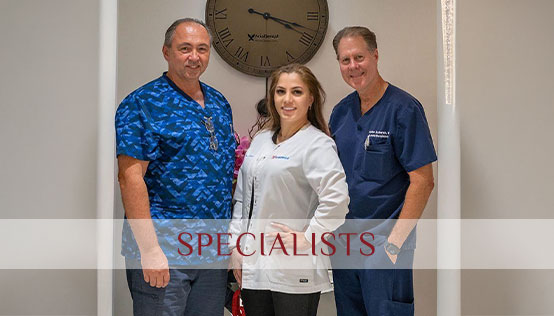
Dr. Horiyat also works alongside a team of experienced dental specialists, so complex cases benefit from coordinated planning in one office.
Keeping Your Gums Healthy After Treatment
- Brush with an extra-soft toothbrush and light pressure, or use an electric brush with a pressure sensor
- Clean between teeth every day with floss or interdental brushes
- Wear your nightguard if you clench or grind
- Keep regular professional cleanings at the interval your dentist recommends
- Avoid smoking and vaping
- Eat nutrient-dense foods; vitamin C, for example, plays a known role in collagen formation
- Ask for recession measurements at each visit so small changes are caught early
Take the Next Step Toward Healthier Gums
Receding gums are common, but they are not something you have to accept. Gingival recession surgery offers proven ways to cover exposed roots, ease sensitivity, strengthen thin gums, and protect your teeth for years to come. The best results begin with an accurate diagnosis, a plan that addresses the cause, and a technique matched to your needs.
If you have noticed longer-looking teeth or sensitivity at the gum line, book your appointment online, contact our office, or call or text (949) 364-9600 to schedule a gum health evaluation with Dr. Horiyat. Aria Dental Care is located at 27652 Crown Valley Pkwy, Mission Viejo, CA 92691, and welcomes patients from Laguna Niguel, Rancho Santa Margarita, Ladera Ranch, San Clemente, Irvine, and throughout Orange County.