


Discovering an unusual white patch inside your mouth can be alarming, especially if you have never heard the word used to describe it before. Leukoplakia is one of the most important oral health conditions for every adult to understand, not because it is always dangerous, but because it demands attention, monitoring, and the right professional guidance. It is a condition where your mouth is sending you a signal that something in your environment, habits, or biology has changed, and that signal deserves to be heard.
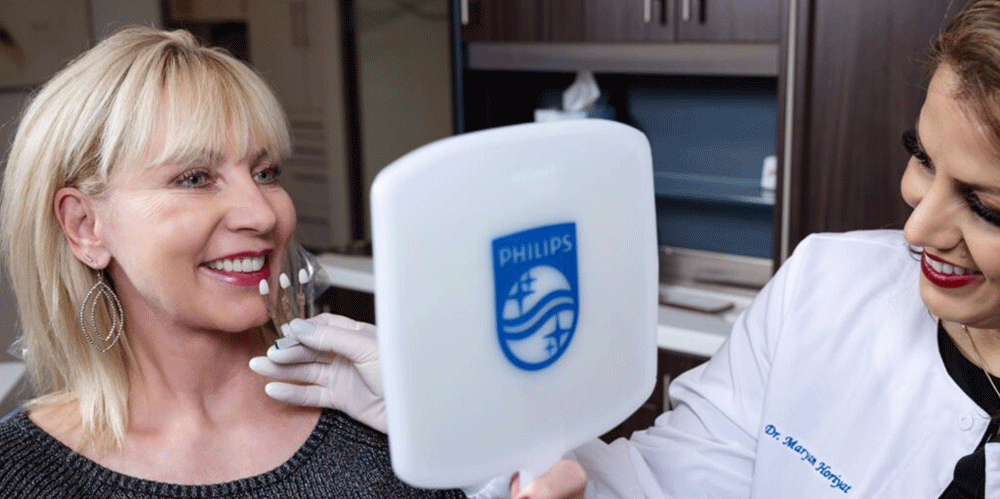
At Aria Dental, Orange County’s #1 Holistic, Biologic & Cosmetic Dental Office, we approach leukoplakia through a whole-body lens. Unlike a purely reactive model of dental care that simply monitors a patch and waits, our holistic philosophy asks a deeper question: why did this develop, and what can we do to address the underlying causes while ensuring your safety? Dr. Maryam Horiyat, DDS, AIAOMT, CIABDM, has extensive training in oral pathology, biological dentistry, and the detection and management of oral lesions, including premalignant oral lesions that require careful, experienced evaluation.
This comprehensive guide covers everything you need to know: what leukoplakia is, what causes it, what it looks like, when it becomes dangerous, how it is diagnosed and treated, how it differs from other white patches in the mouth, and what a truly holistic approach to this condition looks like. Whether you have already been told you have leukoplakia or you simply noticed something unfamiliar in your mouth and want to understand it better, this guide is for you.
What Is Leukoplakia? A Clear, Evidence-Based Definition
Leukoplakia is a clinical term used to describe a white or grayish patch or plaque that develops on the mucous membranes inside the mouth, most commonly on the tongue, the inside lining of the cheeks (buccal mucosa), the gums, the floor of the mouth, or the lips. The word itself comes from Greek: leukos meaning ‘white’ and plakia meaning ‘plaque.’
What makes leukoplakia medically significant is not simply its appearance, but its classification as a potentially premalignant oral lesion. According to the World Health Organization (WHO), leukoplakia is defined as “a white plaque of questionable risk having excluded other known diseases or disorders that carry no increased risk for cancer.” In other words, it is a diagnosis of exclusion, a lesion is classified as leukoplakia once other identifiable causes (such as fungal infection or trauma) have been ruled out.
The critical reason leukoplakia requires professional attention is that a proportion of cases, estimated between 1% and 17.5% depending on the subtype, can undergo malignant transformation and develop into oral squamous cell carcinoma (oral cancer). This does not mean that every case of leukoplakia is cancer, or that it will necessarily become cancer. The vast majority of lesions remain benign or resolve with appropriate intervention. But the risk is real enough that no case of leukoplakia should be ignored, self-diagnosed, or left without proper professional evaluation.
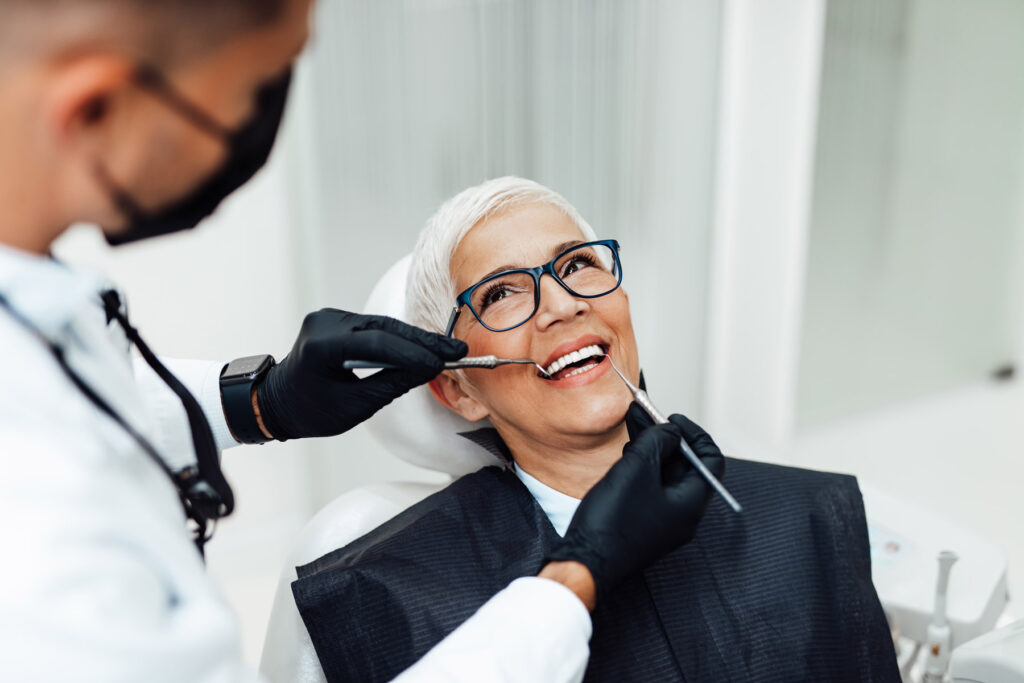
At Aria Dental, we perform comprehensive oral lesion screenings at every routine dental visit, because early detection of leukoplakia and other oral abnormalities is one of the most powerful tools we have for protecting your long-term health.

Types of Leukoplakia: Understanding the Spectrum from Homogeneous to Erythroleukoplakia
Homogeneous Leukoplakia
Homogeneous leukoplakia is the most common and generally the least concerning subtype. It presents as a uniformly white, flat or slightly raised patch with a consistent texture and well-defined borders. It may have a slightly wrinkled or smooth surface but remains consistent in color throughout. Homogeneous leukoplakia carries a relatively lower risk of malignant transformation, though it still requires regular monitoring.
Non-Homogeneous Leukoplakia
Non-homogeneous leukoplakia is the subtype that warrants the greatest clinical concern. It presents with an irregular surface texture and may include nodular (bumpy), verrucous (warty), or ulcerated areas. The color may vary, mixing white with red areas, and the borders may be less well-defined. Non-homogeneous lesions carry a significantly higher risk of malignant transformation and require prompt biopsy and close follow-up.
Erythroleukoplakia (Speckled Leukoplakia)
Erythroleukoplakia, also called speckled leukoplakia or erythroplakia with leukoplakic components, is characterized by a mix of white and red areas within the same lesion. It is considered one of the highest-risk subtypes of oral mucosal lesions. Studies suggest that erythroleukoplakia has a malignant transformation rate significantly higher than purely white lesions, making immediate evaluation and biopsy essential.
Proliferative Verrucous Leukoplakia (PVL)
Proliferative verrucous leukoplakia is a particularly aggressive and challenging subtype. It tends to begin as a flat white patch but gradually develops a thick, warty, multifocal surface over time. It is more common in older women who are non-smokers, an epidemiological profile that makes it easy to misclassify or dismiss. PVL has a very high rate of malignant transformation (up to 70% in some studies) and requires ongoing, vigilant long-term monitoring by an experienced clinician.
What Causes Leukoplakia? Risk Factors and Triggers Explained
Understanding what causes leukoplakia is essential both for prevention and for the holistic management philosophy we practice at Aria Dental. Leukoplakia is not random, it develops in response to identifiable stressors, irritants, and systemic factors. Here are the primary causes and risk factors:
Tobacco Use: The Most Significant Modifiable Risk Factor
Tobacco use, in all its forms, is the single most important risk factor for leukoplakia and the most actionable one. Cigarette smoking, cigar and pipe smoking, chewing tobacco, snuff, and betel quid all create chronic chemical and thermal irritation to the oral mucosa that triggers abnormal keratinization of the tissue lining the mouth.
The carcinogens present in tobacco products do not merely irritate the mucosa, they cause direct DNA damage in oral epithelial cells, which over time can lead to dysplasia (abnormal cell growth) and, ultimately, malignant transformation. The relationship between tobacco and leukoplakia is dose-dependent: the longer and more heavily you use tobacco, the greater your risk. Cessation of tobacco use is one of the most effective interventions for resolving or stabilizing leukoplakia, and it remains a cornerstone of our holistic patient counseling at Aria Dental.
Alcohol Consumption
Chronic and heavy alcohol consumption is an independent risk factor for leukoplakia and oral cancer, and its effects are multiplicative when combined with tobacco. Alcohol acts as a solvent that increases the permeability of oral mucosa to carcinogens, and acetaldehyde (a metabolic byproduct of alcohol) is itself a known carcinogen. Patients who both smoke and drink heavily have a dramatically elevated lifetime risk compared to those with either risk factor alone.
Chronic Mechanical Irritation
Chronic, low-grade mechanical irritation from rough or broken dental restorations, ill-fitting dentures, jagged teeth, or habitual cheek biting can create localized areas of mucosal thickening and white plaque formation. From a holistic dentistry perspective, these are among the most preventable causes of leukoplakia, and addressing them promptly is a core component of our preventive care philosophy at Aria Dental. A comprehensive dental examination identifies sharp edges, poorly fitting appliances, and occlusal issues that contribute to chronic mucosal irritation.
Human Papillomavirus (HPV)
Certain strains of human papillomavirus (HPV), most notably HPV-16, have been identified in a subset of oral leukoplakia cases, particularly those with higher malignant potential. The relationship between HPV and oral cavity lesions mirrors its established role in oropharyngeal cancer and cervical dysplasia. While HPV-associated leukoplakia is less common than tobacco-related cases, its identification has important implications for prognosis and management.
Sun Exposure
Chronic sun exposure is a significant risk factor specifically for leukoplakia of the vermilion border (the junction of the lip and facial skin), particularly the lower lip. Actinic cheilitis, a related premalignant condition of the lip caused by UV radiation, can present with features overlapping those of leukoplakia. Patients who work outdoors or live in sunny climates should use SPF lip balm consistently and have their lips examined at every dental visit.
Nutritional Deficiencies and Systemic Factors
From a holistic and biological dentistry perspective, the role of nutritional status in oral mucosal health is profoundly important. Deficiencies in iron, vitamin B12, folate, and vitamin A have been associated with impaired mucosal integrity and increased susceptibility to oral lesions. Immunosuppression, whether from systemic disease, medications, or chronic stress, reduces the body’s ability to mount appropriate responses to cellular damage and infection, creating conditions in which abnormal tissue changes are more likely to progress unchecked.
At Aria Dental, our whole-body approach means we look beyond the lesion itself to assess the systemic context in which it developed. Nutritional support, immune health, and stress management are all part of the broader conversation we have with patients navigating leukoplakia.
What Does Leukoplakia Look Like? Signs and Symptoms to Know
Leukoplakia is often discovered incidentally, by a dentist during a routine examination, because it is most commonly painless and does not cause obvious symptoms in its early stages. This is precisely why regular dental checkups are so critical for early detection.
The visual characteristics of leukoplakia include:
- Color: White, grayish-white, or mixed white and red (in erythroleukoplakia)
- Texture: Flat and smooth in homogeneous cases; thick, rough, nodular, verrucous, or ulcerated in non-homogeneous or proliferative types
- Size: Ranges from a few millimeters to several centimeters; can be localized or multifocal
- Location: Most commonly found on the buccal mucosa (inner cheeks), lateral border and ventral surface of the tongue, floor of the mouth, gingiva (gums), and lip vermilion
- Borders: May be well-defined or irregular depending on subtype• Removability: Cannot be wiped away, this is a key distinguishing feature from oral thrush (candidiasis), which wipes off easily
The floor of the mouth and the ventral and lateral tongue are considered particularly high-risk sites for malignant transformation, lesions in these locations warrant especially prompt evaluation regardless of their visual appearance.
Symptoms that should prompt immediate professional evaluation include:
- Any white or red patch in the mouth that has been present for more than two weeks
- A lesion that increases in size, changes in appearance, or develops a rough or ulcerated surface
- Numbness, pain, or burning in or around a lesion
- Difficulty chewing, swallowing, or moving the tongue or jaw
- Unexplained bleeding from a mucosal surface

Leukoplakia vs Oral Thrush: How to Tell the Difference
One of the most common sources of patient anxiety is confusion between leukoplakia and oral thrush (oral candidiasis), another condition that produces white patches inside the mouth. While both share a similar appearance at first glance, they are entirely different conditions with different causes, risk levels, and treatments. Here is how they compare:
| Leukoplakia | Oral Thrush (Candidiasis) |
| Cannot be wiped off | Wipes off easily, leaving raw tissue |
| Usually painless | Often causes soreness or burning |
| Caused by chronic irritation / tobacco | Caused by Candida yeast overgrowth |
| Requires biopsy to rule out dysplasia | Resolves with antifungal treatment |
| Potential premalignant condition | Benign fungal infection |
The most reliable and definitive way to distinguish leukoplakia from thrush and other white oral lesions is a professional examination, and, when indicated, a tissue biopsy. At Aria Dental, we perform thorough oral lesion assessments at every visit and refer promptly for biopsy when clinical findings warrant it.
Other conditions that can cause white patches in the mouth and must be distinguished from leukoplakia include:
- Lichen planus: An inflammatory autoimmune condition that produces lacy white lines (Wickham’s striae) or erosive areas on the buccal mucosa; generally bilateral and symmetrical
- Fordyce spots: Harmless ectopic sebaceous glands that appear as pale yellow-white spots, most commonly on the inner cheeks and lip mucosa
- Geographic tongue (benign migratory glossitis): A benign inflammatory condition of the tongue producing map-like white borders surrounding smooth red patches
- Leukoedema: A common, entirely benign whitish film of the buccal mucosa that disappears when the tissue is stretched, unlike leukoplakia, which remains visible
- White sponge nevus: A rare hereditary condition producing white, spongy mucosal plaques
How Is Leukoplakia Diagnosed? The Evaluation Process at Aria Dental
Diagnosing leukoplakia is a process, not a single test. Because it is a diagnosis of exclusion, the clinical pathway involves systematic evaluation to rule out other causes before the leukoplakia designation is confirmed, and to stratify the risk of malignant transformation once confirmed. Here is how that process unfolds at Aria Dental:
Step 1: Comprehensive Visual Oral Examination
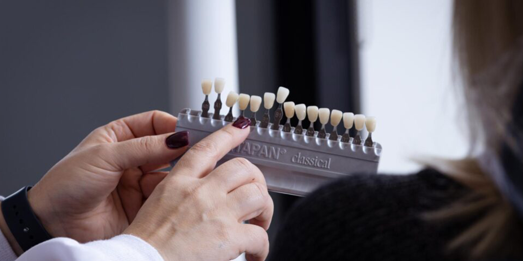
Every routine dental visit at Aria Dental includes a thorough visual and tactile examination of all oral mucosal surfaces, including the tongue, floor of the mouth, palate, cheeks, lips, and gums. We look for any unusual changes in color, texture, contour, or symmetry. If a lesion is identified, we document its size, location, characteristics, and any symptoms the patient reports.
Step 2: Medical and Lifestyle History
Understanding the full context of a lesion is essential. Dr. Horiyat will review your tobacco and alcohol use history, any medications you are taking (some can cause mucosal changes), your nutritional status, immune health, history of HPV, dental appliance fit, and any recent trauma or changes in oral hygiene habits. This comprehensive history is what allows us to identify the most likely contributing factors and recommend the most appropriate initial management.
Step 3: Elimination of Potential Causes
If mechanical irritation from a sharp tooth edge or ill-fitting appliance is suspected, the irritant is removed and the lesion is monitored for resolution over two to four weeks. If a lesion resolves after removing the suspected cause, it may not represent true leukoplakia. If it persists, or if no obvious irritant can be identified, further evaluation is indicated.
Step 4: Adjunctive Screening Technologies
At Aria Dental, we utilize advanced adjunctive oral cancer screening technologies to enhance our ability to identify areas of mucosal abnormality that may not be visible to the naked eye. These tools, including fluorescence-based visualization devices, can help identify areas of tissue that may warrant biopsy even before clinical suspicion is high. While these technologies do not replace biopsy, they provide an additional layer of protection for our patients.
Step 5: Biopsy and Histopathological Analysis
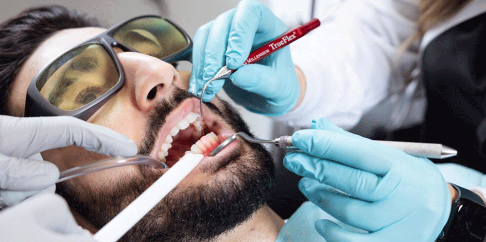
The definitive diagnosis of leukoplakia and the assessment of dysplasia (abnormal cellular changes) can only be made through tissue biopsy and histopathological analysis by an oral pathologist. A biopsy involves removing a small sample of tissue from the lesion under local anesthesia, a minor procedure that takes only minutes. The sample is then examined under a microscope to determine whether dysplastic changes are present and, if so, their severity (mild, moderate, or severe dysplasia).
The degree of dysplasia is the single most important prognostic factor in leukoplakia. Lesions with severe dysplasia or carcinoma in situ require immediate surgical management, while those with mild dysplasia may be managed more conservatively with close monitoring and lifestyle modification.
According to the National Cancer Institute, approximately 3 to 17.5 percent of leukoplakia cases will transform into oral squamous cell carcinoma over a ten-year period, with non-homogeneous lesions and those with high-grade dysplasia carrying significantly greater risk.
Leukoplakia Treatment Options: From Lifestyle Changes to Surgical Intervention
The treatment of leukoplakia is individualized based on the lesion’s characteristics, its degree of dysplasia, its location, the patient’s overall health, and, at Aria Dental, the patient’s lifestyle, nutritional status, and whole-body health context. Here is an overview of the full spectrum of management options:
Tobacco and Alcohol Cessation: The Single Most Impactful Intervention
For tobacco-associated leukoplakia, cessation is not optional, it is the most important intervention available. Studies demonstrate that a significant proportion of leukoplakia lesions, particularly homogeneous, low-dysplasia cases in patients who stop using tobacco, will regress or resolve completely following cessation. For patients who continue to use tobacco, lesions are significantly more likely to persist, progress, and undergo malignant transformation.
At Aria Dental, we provide compassionate, non-judgmental support for tobacco cessation and can connect patients with appropriate resources including cessation counseling and nicotine replacement strategies. We view cessation not as a moral issue but as a medical imperative, one of the most powerful acts of self-care a patient can take.
Elimination of Mechanical Irritants
Where sharp tooth edges, broken restorations, or poorly fitting dentures are contributing to leukoplakia, their prompt correction is both a treatment and a prevention strategy. At Aria Dental, our comprehensive approach to restorative and prosthetic dentistry ensures that every restoration we place and every appliance we fit is smooth, properly contoured, and gentle on oral tissues. This attention to detail is a fundamental expression of our biological dentistry philosophy.
Nutritional and Systemic Support
From a holistic perspective, supporting the body’s own healing capacity is always part of the treatment conversation. Deficiencies in vitamins A, C, E, and B-complex vitamins, as well as iron and zinc, have been associated with impaired mucosal healing and increased dysplasia risk. At Aria Dental, we discuss nutritional optimization as an adjunct to all other treatments, not as a replacement for conventional management, but as a meaningful supportive strategy rooted in biology.
Surgical Excision
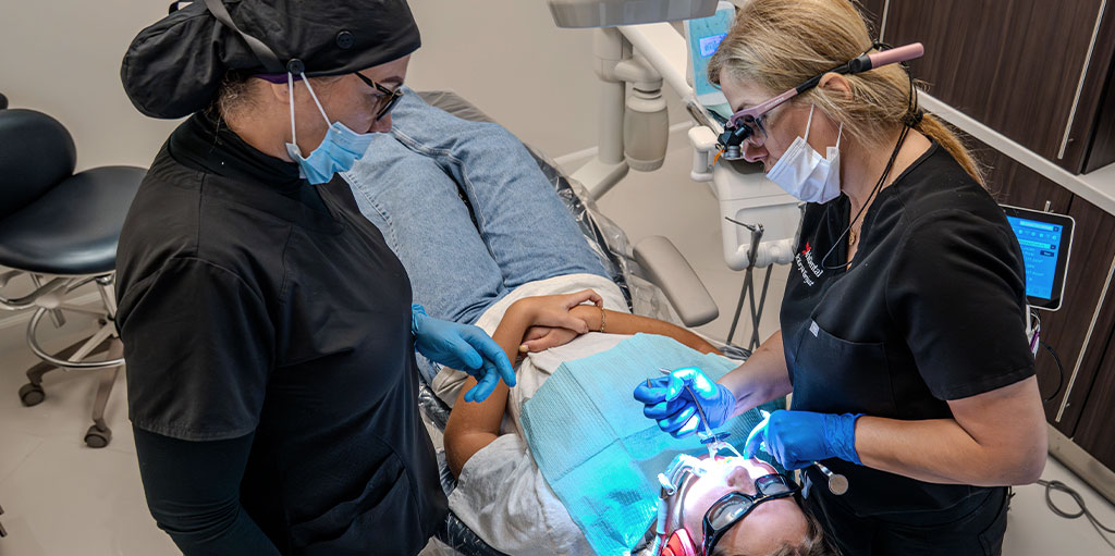
Lesions with moderate to severe dysplasia, high-risk clinical features (non-homogeneous appearance, high-risk site, large size), or confirmed carcinoma in situ require surgical removal. Surgical excision provides both definitive treatment and the most complete histopathological specimen for pathological analysis. It is performed under local anesthesia in most cases and may be done by an oral surgeon or a skilled general dentist with surgical training.
Laser Ablation
Laser ablation, using CO2 or diode laser energy to precisely vaporize abnormal mucosal tissue, has become an increasingly favored treatment modality for leukoplakia, particularly for lesions that are difficult to excise surgically due to size or location. Laser treatment offers excellent precision, reduced bleeding, faster healing, and minimal scarring compared to traditional surgical techniques. At Aria Dental, we utilize state-of-the-art laser technology for soft tissue procedures, consistent with our commitment to offering the latest and most effective dental solutions.
Photodynamic Therapy (PDT)
Photodynamic therapy is an emerging treatment modality in which a photosensitizing agent is applied to the lesion and then activated by a specific wavelength of light, selectively destroying abnormal cells while preserving surrounding healthy tissue. PDT has shown promising results in clinical studies for the management of oral premalignant lesions, including leukoplakia, and represents an exciting frontier in minimally invasive oral medicine.
Long-Term Surveillance
Regardless of the initial treatment chosen, long-term surveillance is essential for all patients with a history of leukoplakia. Even after complete apparent resolution, there is a risk of recurrence, particularly in patients who continue to use tobacco or alcohol. At Aria Dental, we establish individualized monitoring schedules for patients with a history of oral lesions, typically ranging from every three to six months depending on the degree of initial dysplasia and the patient’s ongoing risk profile.
A Holistic Dentist’s Perspective on Leukoplakia: Treating the Whole Person, Not Just the Patch
Conventional dental management of leukoplakia often focuses exclusively on the lesion itself, monitor, biopsy if indicated, excise if dysplastic, and repeat. At Aria Dental, we do all of that, but we also go further, because we believe that a white patch in the mouth is rarely an isolated event. It is a message from the body that the environment in which the oral tissues are living has become inhospitable to healthy function.
Our holistic approach to leukoplakia integrates:
- Root cause identification: What chronic stressors, tobacco, alcohol, nutritional deficiency, immune compromise, poor-fitting appliances, chronic stress, are contributing to this lesion? Can we address them directly?
- Biocompatible oral environment: Are there mercury amalgam restorations creating chronic low-level toxicity in the oral environment? Are there galvanic currents from mixed metals contributing to mucosal irritation? At Aria Dental, our mercury-free, metal-free philosophy minimizes these potential contributors.
- Immune and nutritional support: Is the patient’s immune system supported well enough to mount effective surveillance against cellular dysplasia? What nutritional interventions might reduce inflammatory burden and support mucosal healing?
- Stress and lifestyle medicine: Chronic psychological stress is an underappreciated contributor to immune suppression and impaired tissue healing. We discuss stress management strategies as part of the comprehensive care conversation.
- Environmental toxin reduction: What exposures, dietary, occupational, or environmental, might be contributing to oxidative stress in the oral tissues?
This integrated, systems-thinking approach to leukoplakia management is not a rejection of evidence-based dentistry, it is an expansion of it. Dr. Horiyat’s training in biological dentistry, oral medicine, and the IAOMT’s framework of whole-body oral health uniquely positions her to offer this level of comprehensive, individualized care.
Preventing Leukoplakia: What You Can Do to Protect Your Oral Health
While not every case of leukoplakia is preventable, particularly those associated with genetic factors or HPV, the majority of cases are strongly linked to modifiable risk factors. Here is what you can do to meaningfully reduce your risk:
- Stop or never start using tobacco in any form, cigarettes, cigars, pipes, chewing tobacco, snuff, or betel quid
- Limit alcohol consumption and avoid combining alcohol and tobacco use
- Attend regular dental checkups at least twice per year to allow early detection of any mucosal changes
- Ensure any sharp, broken, or rough dental restorations are promptly smoothed or repaired
- Have ill-fitting dentures or dental appliances adjusted or remade to prevent chronic mucosal trauma
- Eat a nutrient-rich, whole-food diet with adequate intake of vitamins A, C, E, B12, and folate, and minerals including zinc and iron
- Apply SPF lip balm daily, particularly if you spend significant time outdoors
- Discuss HPV vaccination with your healthcare provider if appropriate for your age and risk profile
- Practice thorough daily oral hygiene including brushing, flossing, and tongue cleaning to reduce bacterial load and inflammatory stimulus
- Inform your dentist of any new mucosal changes promptly rather than waiting for your next scheduled visit
At Aria Dental, oral cancer screening and mucosal assessment are not optional add-ons, they are integrated into every routine examination because we know that early detection saves lives.
Protect Your Oral Health with Aria Dental’s Holistic Approach
Leukoplakia is a condition that asks something important of both patient and provider: attention, thoroughness, and a genuine commitment to understanding the whole person behind the lesion. At Aria Dental, we do not just examine your teeth, we examine your oral health in the full context of your life, your biology, and your overall well-being.Dr. Maryam Horiyat, DDS, AIAOMT, CIABDM, brings an exceptional depth of training in biological and holistic dentistry, oral pathology screening, laser soft tissue therapy, and whole-body oral medicine. Our state-of-the-art practice in Orange County is equipped with the latest adjunctive screening technologies, the most biocompatible treatment materials, and a patient-first philosophy that puts your safety, comfort, and long-term health above everything else.Whether you have noticed an unusual patch in your mouth, have been told you have leukoplakia and want a second opinion, or simply want to know that your next routine dental visit includes a thorough oral cancer and mucosal screening, Aria Dental is here for you.
Visit us at ariadentalcare.com to request your comprehensive oral health examination today. Your mouth is telling you something, let us help you listen.